keratocyst

Tratamiento actual en Chile de los queratoquistes odontogénicos
Also called keratocystic odontogenic tumor.
Benign cyst with aggressive behavior.
- Queratoquiste maxilar
- Queratoquiste mandibular
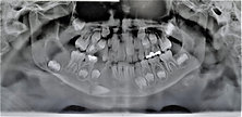
This injury is highly relevant for different reasons. It usually presents without symptoms and can be detected by routine X-rays.
If we look at the radiograph on the left, we can see circular areas in some parts of the mandible. Those are jaw cysts.
The definitive diagnosis is obtained with a biopsy, the moment and type of treatment must be analyzed by your team of surgeons.
Diagnóstico de un queratoquíste
El diagnóstico de un queratoquiste maxilar o mandibular se realiza combinando la presentación clínica y radiológica con el resultado definitivo que entrega una biopsia de la zona que será estudiada por un patólogo maxilofacial.
En nuestra clínica contamos con los protocolos más actuales disponibles para el diagnóstico. contamos con una gran experiencia y un número de casos en control y seguimiento de su tratamiento por muchos años.
Tratamiento de un queratoquiste
El tratamiento tiene básicamente tres etapas.
- Fase 1: La primera conste en la toma de biopsia y descompresión, instalamos una cánula descompresiva y tomamos la muestra para su biopsia.
- Fase 2: Confirmado el diagnóstico por el patólogo realizamos un seguimi8ento y control de su tamaño, luego de seis meses el quiste esté en condiciones de pasar a la tercera fase de tratamiento.
- Fase 3. Extirpación propiamente tal: En este momento realizamos la cirugía para retirar la mayor parte de la membrana del quiste y aplicamos un medicamento que tiene como función frenar el crecimiento celular. El medicamento se llama 5-fluoracilo.
- Fase 4: Seguimiento y controles. Esta fase es clave dado que los queratoquistes tienen una alta posibilidad de regresar, es fundamental un seguimiento e intercepción temprana de la aparición de focos de crecimiento.
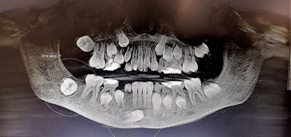
We show a case of a 9-year-old boy in whom different areas with areas of lower bone density were detected by means of an X-ray. In these areas we carry out the elimination through surgery and after a year we can see in the following X-ray that the definitive teeth are being located in their positions and that the areas of the cysts are no longer observed.
On the left side of your screen you can see a large darker area on the molar and in the central part on the tooth that is displaced downwards a similar image.
The areas that presented keratocysts after one year are filled with formation of bone tissue.
Timely treatment is key to a favorable outcome.
Dr. Antonio Marino
Maxillofacial surgeon